Contents
Overview
The study of microscopic anatomy traces its roots back to the invention of the microscope. Early microscopists like Antonie van Leeuwenhoek meticulously documented single-celled organisms and cellular structures, laying the groundwork for a systematic understanding of biological tissues. By the 19th century, figures began to classify tissues based on their physical properties, establishing the concept of distinct tissue types. The formalization of histology as a distinct discipline accelerated with advancements in staining techniques and lens technology, enabling researchers to differentiate cellular components and extracellular matrices with unprecedented clarity. This era saw the rise of dedicated histology textbooks and university courses, solidifying its place in the scientific curriculum.
⚙️ How It Works
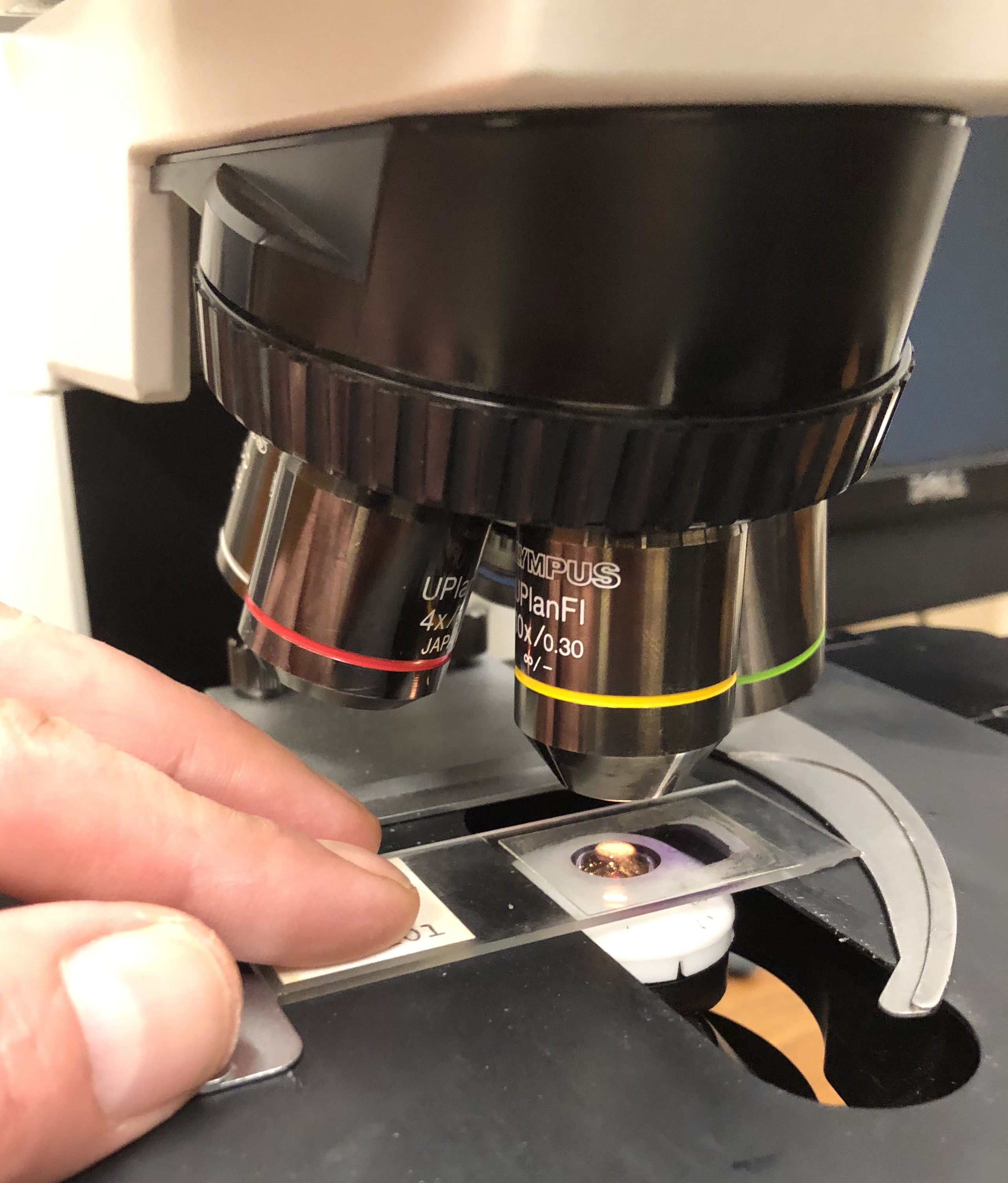
Microscopic anatomy operates by preparing biological specimens for examination under a microscope, a process that typically involves several critical steps. Tissues are first fixed, often with formalin, to preserve their structure and prevent degradation. They are then embedded in a supporting medium, such as paraffin wax or epoxy resin, allowing for the creation of extremely thin sections using a microtome. These thin sections are mounted on glass slides and subjected to various staining procedures, with hematoxylin and eosin (H&E) being the most common, to highlight different cellular and extracellular components. Specialized stains, like Masson's trichrome or Periodic Acid-Schiff (PAS), are used to emphasize specific structures like collagen or carbohydrates, respectively. Finally, these stained slides are viewed under light microscopes, or increasingly, electron microscopes for ultrastructural detail.
📊 Key Facts & Numbers
The field of microscopic anatomy relies on precise measurements and quantifiable data. Transmission electron microscopes (TEMs) can reveal details like ribosomes and endoplasmic reticulum. Neurons are among the longest cells in the body, sometimes extending over a meter. Histopathology reports, crucial for cancer diagnosis, analyze tissue samples. Understanding these microscopic details is crucial for biological and medical advancements.
👥 Key People & Organizations
Key figures in the development of microscopic anatomy include Robert Hooke, credited with coining the term 'cell'. Antonie van Leeuwenhoek, a Dutch draper and scientist, independently developed powerful single-lens microscopes and was the first to observe and describe bacteria, protozoa, and sperm cells. Figures began to classify tissues based on their physical properties. In the 20th century, advancements in electron microscopy were spearheaded by scientists like Ernst Ruska, who co-invented the TEM. Leading institutions like the University of Cambridge and Johns Hopkins University have historically been centers for histological research and education.
🌍 Cultural Impact & Influence
Microscopic anatomy has profoundly shaped our understanding of life and disease, influencing fields from medicine to evolutionary biology. The ability to visualize cellular and tissue structures has been pivotal in identifying the cellular basis of numerous diseases, transforming pathology from a macroscopic to a microscopic discipline. This detailed understanding has directly led to the development of targeted therapies, such as chemotherapy drugs that exploit differences in cancer cell proliferation rates. Furthermore, the study of fossilized tissues, or paleohistology, provides critical insights into the evolution of life, allowing scientists to infer the physiology and lifestyle of extinct organisms, such as the vascularization patterns in dinosaur bones. The visual language of histology, with its stained slides and cellular diagrams, has also permeated scientific illustration and educational materials, becoming an iconic representation of biological inquiry.
⚡ Current State & Latest Developments
The field of microscopic anatomy is currently experiencing a renaissance driven by technological innovation and interdisciplinary collaboration. Digital pathology is rapidly transforming the field, with high-resolution whole-slide imaging allowing for remote consultation, AI-driven analysis, and quantitative measurements that were previously impossible. Advances in immunohistochemistry and fluorescence microscopy enable researchers to visualize specific proteins and molecules within their cellular context, leading to breakthroughs in understanding cellular signaling pathways and disease mechanisms. The integration of genomics and proteomics with traditional histology, often termed 'spatial biology,' is providing unprecedented insights into how gene expression and protein function vary across different cell types and tissue microenvironments. The development of new imaging modalities, such as confocal microscopy and super-resolution microscopy, continues to push the boundaries of what can be visualized at the molecular level.
🤔 Controversies & Debates
While microscopic anatomy is a well-established science, debates persist regarding the interpretation of certain cellular structures and their precise functions, particularly in complex disease states. A significant ongoing discussion revolves around the role of the extracellular matrix in cell signaling and tissue remodeling, with some researchers emphasizing its passive structural role while others highlight its active participation in cellular behavior. The classification of certain cell types or tissue subtypes also remains a point of contention, especially in oncology, where subtle morphological differences can have profound prognostic implications. Furthermore, the ethical considerations surrounding the use of human tissue samples for research, particularly concerning donor consent and data privacy in the era of digital pathology and AI analysis, are subjects of continuous debate within the scientific and medical communities.
🔮 Future Outlook & Predictions
The future of microscopic anatomy is inextricably linked to advancements in imaging technology, computational biology, and artificial intelligence. We can anticipate the development of even higher resolution microscopes capable of visualizing molecular interactions in real-time within living tissues, a field known as live-cell imaging. The integration of AI in digital pathology is poised to revolutionize diagnostics, potentially identifying subtle patterns indicative of disease far earlier and more accurately than human pathologists alone. The concept of 'digital twins' for organs or even entire patients, built from detailed histological and molecular data, could enable personalized treatment planning and drug development. Furthermore, the application of microscopic anatomical principles to fields like bioengineering and tissue engineering will likely lead to the creation of more sophisticated artificial organs and regenerative therapies, blurring the lines between biological and synthetic systems.
💡 Practical Applications
Microscopic anatomy finds critical applications in numerous fields. In medicine, histopathology is essential for diagnosing diseases, particularly cancers, by examining tissue samples for abnormalities. It guides treatment decisions and monitors disease progression. In research, it's fundamental for understanding normal biological function and the mechanisms of disease at the cellular level. Forensic science utilizes microscopic analysis of tissues to aid in investigations. In dentistry, it's used to study the microscopic structure of teeth and surrounding tissues. The field also supports the development of new drugs and therapies by providing insights into cellular targets and responses.
Key Facts
- Category
- science
- Type
- topic